Cellular Reprogramming Technologies for Disease Modeling
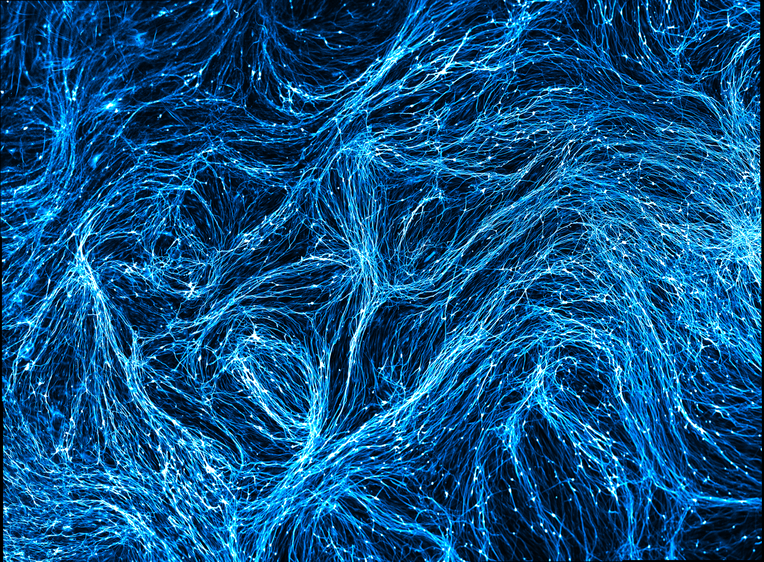
During my graduate training, I developed a cellular reprogramming method to generate striatal medium spiny neurons (MSNs), the primary cell type affected in Huntington's disease (HD). This approach takes advantage of the synergistic activity of brain-enriched microRNAs with transcription factors expressed in the developing striatum to bypass the induction of pluripotency and directly convert human adult fibroblasts into neurons. During the course of my thesis, we found that unlike iPSC-based methods, directly converted MSNs retain the phenotypic age of the starting adult fibroblasts. We hypothesized that directly converted MSNs from HD patients could offer advantages in modeling HD in tissue culture. In HD, mutant Huntingtin protein (mHTT) can misfold and aggregate in the brain, eventually leading to degeneration of MSNs. Directly converted MSNs generated from HD patient fibroblasts exhibited mHTT aggregates, and spontaneously degenerated over time in culture. This was a particularly striking observation given that MSNs generated through iPSC-based methods lack overt phenotypes. I discovered that differences in mHTT aggregation propensity in direct conversion versus iPSC-based methods are the result of drastically different levels of proteostasis conferred by these two distinct reprogramming methods. While the induction of pluripotency promotes cellular rejuvenation, directly converted cells retain the age-associated decline in proteosome activity levels. Our findings address many of the challenges faced by modeling late-onset diseases using neurons differentiated from iPSCs while establishing a novel human cellular platform to screen potential disease-halting interventions for HD.
Victor M.B., Richner M., Hermanstyne T.O., Ransdell J.L., Sobieski C., Deng P.Y., Klyachko V.A., Nerbonne J.M., Yoo A.S. Generation of Human Striatal Neurons by MicroRNA-Dependent Direct Conversion of Fibroblasts. Neuron (2014); 84 (2): 311–323.
Richner M., Victor M.B., Liu Y., Abernathy D.G., Yoo A.S. MiRNA-based Conversion of Human Fibroblasts to Striatal Medium Spiny Neurons. Nature Protocols (2015); 10(10):1543-55.
Huh C.J., Zhang B., Victor M.B., Dahiya S., Batista L.F.Z., Horvath S., Yoo A.S. Maintenance of Age in Human Neurons Generate by microRNA-based Neuronal Conversion of Fibroblasts. eLife (2016);5:e18648
Victor M.B., Richner M., Olsen H., Lee S., Monteys M.A., Ma C., Huh C., Zhang B., Davidson B.L.,Yang X.W. & Yoo A.S. Modeling Huntington's Disease with Directly Converted Patient Neurons. Nature Neuroscience (2018); 21(3):341-352

Modeling the Bidirectional Communication of Neurons and Microglia
As a postdoctoral fellow in the Tsai lab, I have been employing CRISPR-edited induced pluripotent stem cells (iPSCs) to dissect the impact of Apolipoprotein E4 (APOE4) in neuron-microglia communication. APOE4 is the greatest known genetic risk factor for developing late-onset Alzheimer’s disease and its expression in microglia is associated with pro-inflammatory states. How the interaction of APOE4 microglia with neurons differs from microglia expressing the disease-neutral allele APOE3 is currently unknown. Our results revealed that APOE4 induces a distinct metabolic program in microglia that is marked by the accumulation of intracellular neutral lipid stores through impaired lipid catabolism. Importantly, this altered lipid-accumulated state shifts microglia away from homeostatic surveillance and renders APOE4 microglia weakly responsive to neuronal activity. Remarkably, unlike APOE3 microglia that support neuronal network activity, co-culture of APOE4 microglia with neurons disrupts the coordinated activity of neuronal ensembles. We identified that through decreased uptake of extracellular fatty acids and lipoproteins, APOE4 microglia disrupts the net flux of lipids which results in decreased neuronal activity via the potentiation of lipid-gated K+ channels. These findings suggest that abnormal neuronal network-level disturbances observed in Alzheimer’s disease patients may in part be triggered by impairment in lipid homeostasis in non-neuronal cells, underscoring a novel therapeutic route to restore circuit function in the diseased brain.
1. Victor M.B., Leary N., Luna X., Meharena H.S., Scannail A.N., Bozzelli P.L., Samaan G., Murdock M.H., Maydell D.V., Effenberger A.H., Cerit O., Wen S.L., Liu L., Welch G., Bonner M. & Tsai L.H. Lipid Accumulation Induced by APOE4 Impairs Microglial Surveillance of Neuronal-Network Activity. Cell Stem Cell (2022); 29(8):1197-1212
2. Blanchard J.W.*, Victor M.B. *, & Tsai L.H. Dissecting the complexities of Alzheimer disease with in vitro models of the human brain. Nature Reviews Neurology (2022); 18(1):25-39. * Equal Contribution.
3. Blanchard J.W., Bula M., Davila-Velderrain J., Akay L.A., Zhu L., Frank A., Victor M.B., Bonner J.M., Mathys H., Lin Y.T., Ko T., Bennett D.A., Cam H.P., Kellis M., Tsai L.H. Reconstruction of the human blood-brain barrier in vitro reveals a pathogenic mechanism of APOE4 in pericytes. Nature Medicine (2020); 26(6):952-963.

Leveraging iPSCs with Single-Cell Profiling to Advance our Understanding of Alzheimer’s Disease
In collaboration with Manolis Kellis’ Lab at MIT CSAIL, we employed single-nucleus sequencing (snRNA-seq) and captured the transcriptomes of 194,000 microglia isolated from 444 human subjects across six brain regions. Of these individuals, 220 displayed varying degrees of Alzheimer’s disease (AD) pathology. Our data revealed the molecular signatures of 12 distinct microglial states, including inflammatory and lipid processing states that were present in significantly increased proportions in AD individuals and positively correlated with levels of amyloid plaque and tau tangle burden. Using transcription factor-driven regulatory networks, we inferred master regulators of microglial states and their transitions. Through enrichment analysis of microglia-like cells (iMGLs) profiled by snRNAseq, our work defines the transcriptional state of iMGLs in relation to human microglial states. We demonstrate that forced expression of transcription factor regulators of microglia homeostasis can induce homeostatic features in iMGLs, and that inhibiting the induction of transcription factors predicted to drive inflammatory states with CRISPRi is sufficient to block transcriptional features of LPS-induced inflammation. Additionally we leveraged iMGLs to recapitulate transcriptional programs that governs microglial response to the phagocytosis of amyloid beta fibrils, uncovering a temporal program of inflammatory microglia state transitions that map to snRNA-seq of aged human brains. Collectively, our study provides a roadmap to design microglia state specific therapies aimed at curbing neuroinflammation and halting Alzheimer’s disease progression.
Sun N.*, Victor M.B.*, Park Y., Xiong X., Scannail A.N., Leary N., Prosper S., Viswanathan S., Luna X., Boix C.A., James B.T., Tanigawa Y., Galani K., Mathys H., Jiang X., Ng A.P., Bennett D.A., Tsai L.H.†, Kellis M.† Human Microglial State Dynamics in Alzheimer’s Disease Progression. Cell (2023).
* Equal Contribution, † Co-Supervised the Work.
Mathys H.*, Boix C.*, Peng, Z.*, Victor M.B., Leary N., Babu, S., Abdelhady G., Jiang X., Ng A.P., Ghafari K., Kunisky A.K., Matero J., Galani K., Logia V.N., Fortier G.E., Lotfi Y., Ivey J.B, Brown H.P., Patel P.R., Chakraborty N., Beaudway J.I., Imhoff E.J., Keeler C.F., McChesney M.M., Patel H.H., Patel S.P., Thai M.T., Bennett D.A., Kellis M.† , Tsai L.H.† Single-cell atlas of the aged human brain reveals cellular and molecular correlates of high cognitive function, dementia, and resilience to Alzheimer’s disease pathology. Cell (2023).
* Equal Contribution, † Co-Supervised the Work.
Xiong X.*, James B.T.*, Boix C.A.*, Park Y., Galani K, Victor M.B., Sun N., Hou L., Ho L.L., Mantero J., Scannail A.N., Mathys H., Bennett D.A., Tsai L.H.†, Kellis M.† Single-cell epigenomic dissection of Alzheimer's disease pinpoints causal genetic variants and reveals epigenome erosion. Cell (2023).
* Equal Contribution, † Co-Supervised the Work.